738 results

- Digital Images
- Online
Monocyte and two red blood cells
University of Edinburgh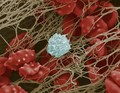
- Digital Images
- Online
Blood clot with crenated red cells
Anne Weston, Francis Crick Institute
- Digital Images
- Online
Red blood cells forming rouleaux
Jonathan Armstrong
- Digital Images
- Online
Red blood cells forming rouleaux
Jonathan Armstrong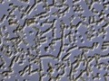
- Digital Images
- Online
Red blood cells forming rouleaux
Jonathan Armstrong- Books
An ode to red blood cells.
Date: [2019]
- Digital Images
- Online
Platelets and red blood cells
University of Edinburgh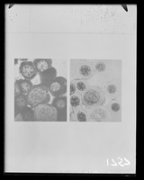
- Archives and manuscripts
- Online
Microscope image referenced as "Blood cells"
Wilkins, Maurice Hugh Frederick, 1916-2004Date: September 1955Reference: KDBP/1/1/1757Part of: King's College London Department of Biophysics
- Archives and manuscripts
- Online
Microscope image referenced as "Blood cells"
Wilkins, Maurice Hugh Frederick, 1916-2004Date: September 1955Reference: KDBP/1/1/1760Part of: King's College London Department of Biophysics
- Archives and manuscripts
- Online
Microscope image referenced as "Blood cells"
Wilkins, Maurice Hugh Frederick, 1916-2004Date: September 1955Reference: KDBP/1/1/1758Part of: King's College London Department of Biophysics
- Digital Images
- Online
Skin around wound, mouse, blood cells
David Gregory & Debbie Marshall
- Digital Images
- Online
Red blood cells clearly showing their biconcave disc shape.
Annie Cavanagh- Books
The morphology of human blood cells / L.W. Diggs, Dorothy Sturm, Ann Bell.
Diggs, L. W. (Lemuel W.), 1900-1995.Date: 1956
- Digital Images
- Online
Red blood cells clearly showing their biconcave disc shape.
Annie Cavanagh- Books
The morphology of blood cells in Wright stained smears of peripheral blood and bone marrow / by L.W. Diggs [and others].
Date: [1954], ©1954- Books
Living blood cells and their ultrastructure / Marcel Bessis ; translated by Robert I. Weed.
Bessis, Marcel, 1917-Date: 1973- Film
Blood flow in arteriovenous anastomoses and deformation of blood cells with increasing velocity.
Date: 196?
- Digital Images
- Online
Trypanosome among blood cells
Gull Lab courtesy of Samantha Griffiths
- Digital Images
- Online
Cellular architecture of normal human skin imaged by whole mount tissue microscopy. Human skin has a rich network of white blood cells (specifically dendritic cells, T cells and macrophages) which form sheaths around blood vessels. In this image, blood vessels (string-like structures stained for CD31; green), lymphatic vessels (ribbon-like structures stained for LYVE-1; blue) and T cells (stained for CD3; red) can be seen. T cells are only found around dermal blood vessels. Macrophages (stained for LYVE-1; blue) are also present. This normal cellular architecture is grossly disrupted in diseased skin (see related images). X10 magnification. Scale bar (white) represents 200 micrometres.
Dr. Xiao-nong Wang, Human Dendritic Cell Laboratory, Newcastle University
- Books
- Online
The biology of the blood-cells : with a glossary of haematological terms; for the use of practitioners of medicine.
Gruner, O. Cameron (Oskar Cameron), 1877-1972.Date: 1913
- Digital Images
- Online
Cellular architecture of normal human skin imaged by whole mount tissue microscopy. Human skin has a rich network of white blood cells (specifically dendritic cells, T cells and macrophages) which form sheaths around blood vessels. In this image, blood vessels (string-like structures stained for CD31; red), lymphatic vessels (ribbon-like structures stained for LYVE-1; blue) and dendritic cells (stained for CD11c; green) can be seen. Macrophages (stained for LYVE-1; blue) are also present. This normal cellular architecture is grossly disrupted in diseased skin (see related images). X10 magnification. Scale bar (white) represents 200 micrometres.
Dr. Xiao-nong Wang, Human Dendritic Cell Laboratory, Newcastle University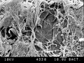
- Digital Images
- Online
SEM of blood clot, new cells under fibrin
David Gregory & Debbie Marshall
- Digital Images
- Online
Cellular architecture of normal human skin imaged by whole mount tissue microscopy. Human skin has a rich network of white blood cells (specifically dendritic cells, T cells and macrophages) which form sheaths around blood vessels. In this image, T cells (stained for CD3; red) dendritic cells (stained for MHC class II; green) and macrophages (stained for LYVE-1; blue with some cells showing a tinge of green) can be seen. Cell nuclei have been stained with DAPI (grey). This normal cellular architecture is grossly disrupted in diseased skin (see related images). X10 magnification. Scale bar (white) represents 200 micrometres.
Dr. Xiao-nong Wang, Human Dendritic Cell Laboratory, Newcastle University
- Digital Images
- Online
Cellular architecture of normal human skin imaged by whole mount tissue microscopy. Human skin has a rich network of white blood cells (specifically dendritic cells, T cells and macrophages) which form sheaths around blood vessels. In this image, T cells (stained for CD3; red) dendritic cells (stained for MHC class II; green) and macrophages (stained for LYVE-1; blue with some cells showing a tinge of green) can be seen. Cell nuclei have been stained with DAPI (grey). This normal cellular architecture is grossly disrupted in diseased skin (see related images). X20 magnification. Scale bar (white) represents 100 micrometres.
Dr. Xiao-nong Wang, Human Dendritic Cell Laboratory, Newcastle University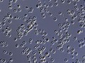
- Digital Images
- Online
Red blood cells from a person with sickle cell anaemia. These cells are not sickled as there is plenty of oxygen present. For a deoxygenated, sickled comparison see N0024943
Jonathan Armstrong